microglia migration assay
However these microglia remained actively moving without any reduction in migration speed. For research questions where it is important that the.

A Schematic Diagram Of The Microglial Migration Assay A Tissue Download Scientific Diagram
Migration assay and additional mechanism studies were performed to elucidate the role of NF-κB in S100B-mediated microglia M1M2 phenotype change and migration.

. Scratch assays were performed following the previously described. For high throughput screening assays the microglial cell lines that are available might suffice using culture conditions with or without serum. While this shows that microglia can respond to ATP regardless of their activation state IL4-treated microglia remained the most migratory and LPS-treated cells the least migratory.
Microglia were seeded on coverslips at 3 10 4 cellsTranswell insert for migration assays 6 10 4 cells15 mm coverslip for fluorescence microscopy and NO production and 10 5 cellscoverslip for mRNA isolation. Bespoke Assay Service In addition to our standard assays Celentyx is often called upon to develop bespoke assays. The secreted factor model uses a 04 μm pore size to exclude microglia cell migration a while the migration model uses an 8 μm pore size to promote microglia cell migration b.
After plating the microglia were incubated for 24 h at which time they were healthy looking see images in Results. After plating the microglia were incubated for 24 h at which time they were healthy looking see images in Results. Microglia were seeded on coverslips at 3 10 4 cellsTranswell insert for migration assays 6 10 4 cells15 mm coverslip for fluorescence microscopy and NO production and 10 5 cellscoverslip for mRNA isolation.
A line down the center of each well was scraped with a sterile p200 pipette tip and followed by a. In combination with microscopic imaging this assay allows simultaneous recording of cell movement and subcellular compartment trafficking along with quantitative analysis. To confirm the role of cathD in microglia migration we next analyzed the directional migration of microglia using an in vitro micropipette assay that mimics endogenous ATP release during injury 18 32.
In the present study we designed an in vitro culture system that allows the study of cell migration across an MBEC monolayer on a Transwell membrane and used this novel assay to define a role for ATP and MMPs in microglia migration across endothelial barriers eg. S5A and movies S13 and 14 as. In the damaged brain additional stimuli and chemotactic.
Briefly the BV-2 microglia in six well plates were performed with serum-free DMEM for three wash times. Compared with wild-type controls cathD-deficient microglia exhibited a suppressed migration activity upon ATP-γ-S stimulation fig. Most commonly used cell culturebased migration assays is the Boyden chamber Transwell assay which consists of an insert with a porous membrane lining nested in a culture plate 1619.
After migration the filter is stained and the cells are counted. The Boyden chamber assay is relatively simple to perform. Chemotactic migration of these microglial cells toward Aβ aggregates was significantly attenuated by TGF-β1.
In response to injury or inflammatory stimuli the resting microglia can be rapidly activated to participate in pathological. It involves the migration of cells through a microporous filter toward a high concentration of chemoattractant placed in a well below the filter. Incucyte Scratch Wound and Chemotaxis Assays allow you to continuously monitor and analyze migration and invasion with or without a chemotactic gradient right inside your incubator.
Migration of bv-2 microglial cells was evaluated using a chemotaxis boyden chamber system with 24-well insert with 80 μm pore size polycarbonate membrane separating upper and lower wells spl life sciences korea. This protocol is an adaptation of the axon turning assay using microglial cells. The assay is long and tedious has poor reproducibility requires a large number of.
Pharmacological blockade of TGF-β1 receptor I ALK5 by SB431542 treatment reduced the inhibitory effects of TGF-β1 on Aβ-induced BV-2 microglial. BV2 cells or primary microglia are seeded onto transwell inserts and then inserted into 24-well plates with differentiated SH-SY5Y cells grown on coated glass coverslips. Microglia cells 1x10⁵ cells in 100 μl 03 BSARPMI1640.
Chemoattractants released from the micropipette tip produce a chemotactic gradient that induces robust microglial migration. Microglia are resident immune cells in the central nervous system CNS. In addition it is of importance that doubts on the species origin of human cell lines will be unequivocally confirmed and if appropriate accepted and corrected for.
S100B was identified as an induced gene with its pattern in accordance with M1 markers in mice MCAO. In the presence of microglia glioma cell migration occurred earlier and after 48 h it was threefold higher as compared to incubations without microglia. Incucyte live-cell assays can be conducted either label-free or by using dual color fluorescence to study specific cell populations in co-culture.
The migration assay was performed with filters coated with midkine MK or poly-D-lysine PDL on the lower surface at 10 μgml. Microglia differentiated from peripheral blood monocytes phagocytosing pH-rodo-labelled myelin basic protein red when intracellular over 14 hrs. We therefore studied the effects of murine microglial cells on motility of GL261 mouse glioma cells using Boyden chamber assays.
Moreover In vitro migration assays scratch assay and transwell migration assay were performed. However it has several limitations. Finally S100B treatment on MCAO models was performed to show the in vivo evidence.
This effect was mediated by substances released from microglia because similar effects were. 1 10 5 cells100 μlwell were seeded into the upper inserts while treatment media was applied to the lower wells and incubated. In all of our assays ATP increased microglia migration whether the cells were untreated or stimulated with IL4 or LPS.

Inflammatory Interactions Between Degenerated Intervertebral Discs And Microglia Implication Of Sphingosine 1 Phosphate Signaling Navone 2021 Journal Of Orthopaedic Research Wiley Online Library

Cxcl10 Triggers Early Microglial Activation In The Cuprizone Model The Journal Of Immunology

A Schematic Diagram Of The Microglial Migration Assay A Tissue Download Scientific Diagram

Curcumin Reduces Microglial Migration A B Scratch Assays In Bv 2 Download Scientific Diagram

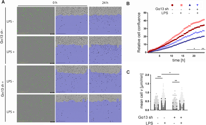
Ga13 Contributes To Lps Induced Morphological Alterations And Affects Migration Of Microglia Springerlink

Dex Sp Inhibited Migration Of Bv 2 Microglia Cell In Vitro The Download Scientific Diagram

Hlp Promotes Microglial Migration A Schematic Representation Of The Download Scientific Diagram

Tlqp21 Affects Microglial Phagocytic Activity A Phagocytosis Assay In Download Scientific Diagram

The Effect Of Different Tn R Domains On Microglia Migration The Download Scientific Diagram

Analysis Of Cell Migration Determined By Boyden Chamber Assay A And Download Scientific Diagram

Gingipains Promote The Cell Migration After The Infection Of Microglia Download Scientific Diagram

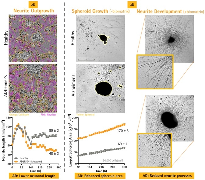
Neuronal And Microglial Analysis For Alzheimer S Disease Models

Curcumin Reduces Microglial Migration A B Scratch Assays In Bv 2 Download Scientific Diagram

Migration And Chemotaxis Are Affected By The Microglial Activation Download Scientific Diagram

Puma Mediated The Methamphetamine Induced Migration Of Microglia A A Download Scientific Diagram

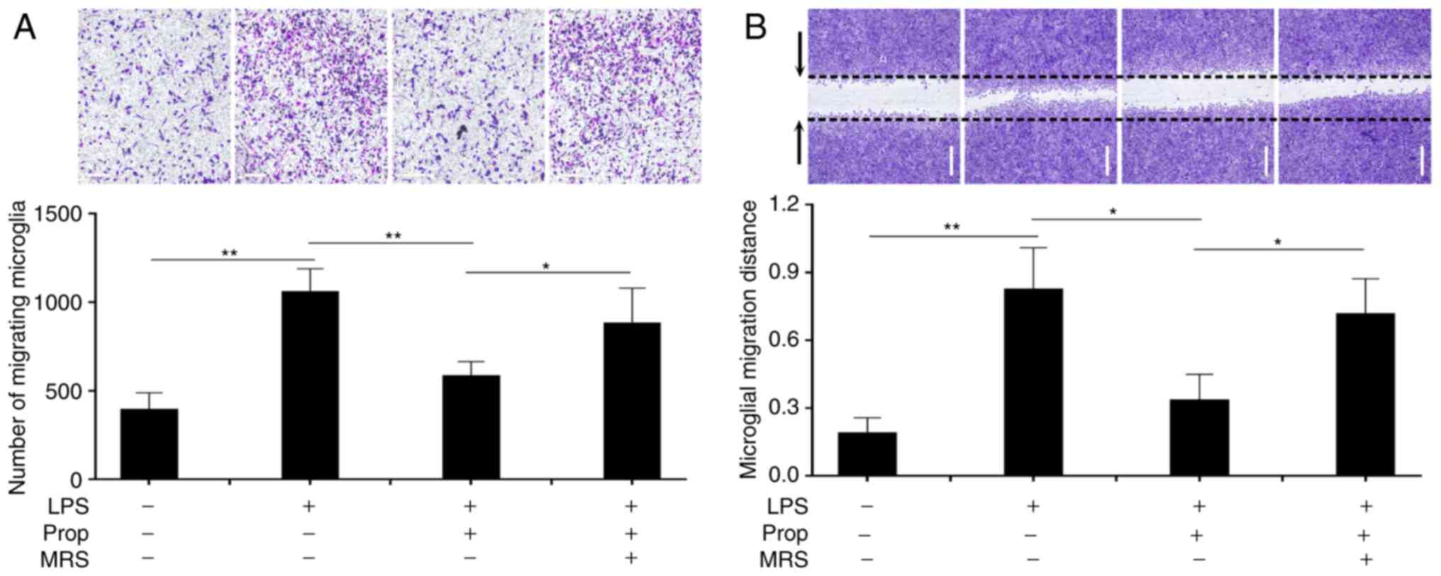
Propofol Attenuates Inflammatory Damage On Neurons Following Cerebral Infarction By Inhibiting Excessive Activation Of Microglia

Jci Insight Integrin Kindlin3 Requirements For Microglial Motility In Vivo Are Distinct From Those For Macrophages

A Schematic Diagram Of The Microglial Migration Assay A Tissue Download Scientific Diagram

Scutellarin Inhibited Migration Of Bv 2 Microglia Transwell Migration Download Scientific Diagram
Comments
Post a Comment